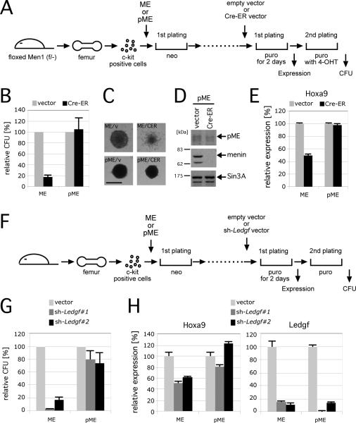
Figure 5. LEDGF is necessary for maintenance of MLL leukemic transformation.
A. The experimental scheme for conditional inactivation of Men1 in MLL transformed cells. The time points at which CFU or gene expression were measured are indicated.
B. The relative CFUs are shown for cells transformed by ME or pME in the absence (vector) or presence (Cre-ER) of Men1 inactivation (vector controls are arbitrarily set at 100%). Error bars represent the standard deviations of three independent analyses.
C. Morphologies are shown for representative colonies from experiment in panel B. Scale bar, 150 μm.
D. Western blot shows expression of pME, menin and SIN3A proteins after Men1 inactivation.
E. Relative expression levels of Hoxa9 are shown for first round colonies after Cre-ER transduction. Expression levels were normalized to β-Actin, and expressed relative to the vector control values (arbitrarily set at 100%). Error bars represent standard deviations of triplicate PCR analyses.
F. Experimental scheme for conditional inactivation of Ledgf by shRNA-mediated knockdown. The time points at which CFUs or gene expression were measured are indicated.
G. The relative CFU activity of ME- or pME-transformed cells is shown with (sh-Ledgf#1 or sh-Ledgf#2) or without (vector) Ledgf knockdown (the vector controls are arbitrarily set at 100%). Error bars represent the standard deviations of three independent analyses.
H. Relative expression levels of Hoxa9 and Ledgf are shown for first round colonies after shRNA vector transduction. Expression levels are normalized to β-actin, and expressed relative to the vector control values (arbitrarily set at 100%). Error bars represent the standard deviations of triplicate PCR analyses.

