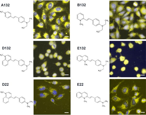
Figure 2.
Images of cells incubated with a group of related styryl probes, exhibiting normal (A132, B132, D22 and E22) and rounded (D132, E132) cell shape phenotypes. All images are composites acquired from the Hoechst™ (blue) and TRITC channel (yellow, 1s exposure with extracellular dye). Scale bar = 10 μm.

