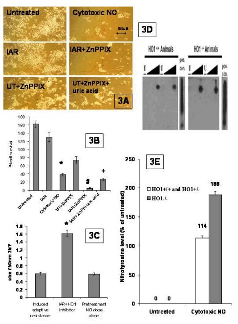
Fig. 3. A more specific HO1 inhibitor abrogates IAR.
A. Micrographs of motor neurons: NSC34 cells were subjected to the IAR protocol(IAR), the IAR protocol in the presence of the HO1 inhibitor (IAR+ZnPP), the untreated cells incubated with ZnPPIX (UT+ZnPPIX), and the IAR in the presence of the HO1 inhibitor and uric acid (IAR + ZnPP + uric acid). (100×magnification) B. Quantification of cell survival and HO1 inhibition: The experiments were repeated, the cell counts were reported, and a comparison between IAR and cytotoxic NO was determined as significant (p<.001) and designated by an asterisk. A comparison between IAR and IAR +ZnPPIX was determined as significant (p<.001) and designated by a #. A comparison between IAR +ZnPPIX and IAR+ ZnPPIX +uric acid was determined to be significant (p<.01) and designated by +. C. ELISA analysis of 3NY levels. 3NY levels in IAR and HO1 inhibited IAR cells were measured and the difference was determined to be significant (p<.001): D. Western of 3NYin HO1 wildtype and HO1 null cells: Primary motor neurons were isolated from wildtype, heterozygous, or HO1 null mice. The groups of cells were untreated (u), or treated with a low NO dose (l) or medium NO dose (m) or high NO dose (h). Protein was isolated and westerns run and probed for nitrotyrosine. E. Quantification of 3NY levels: The protein levels were checked for even loading and nitrotyrosine levels were quantified against controls by densitometry. This was repeated, averaged, an SEM was calculated, and significance determined.

