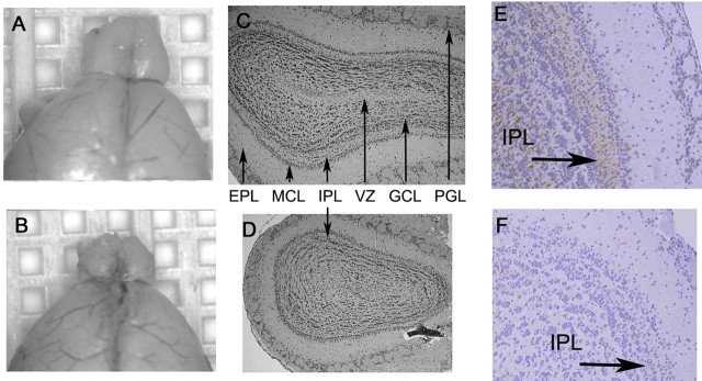
Figure 1.
Id2−/− mice have a diminished olfactory bulb. A, B, Micrographs of representative gross morphology of WT (A) and Id2−/− (B) brains. C, D, Morphometrically aligned coronal sections stained with hematoxylin and eosin [arrows denote PGL, external plexiform layer (EPL), mitral cell layer (MCL), IPL, granular cell layer (GCL), and ventricular zone (VZ)]. E, F, Immunohistochemical evaluation of OB histologic sections from WT (E) and Id2−/− (F) mice for neurofilament expression visualized by brown DAB staining and hematoxylin counterstain. Arrows indicate location of IPL, original magnification 40×.

