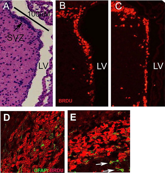
Figure 4.

Id2−/− mice retain normal numbers of neural progenitor cells, but the RMS is reduced in size and disorganized. A–C, Multiple sections taken from short-term BrdU pulsed animals were immunostained to identify BrdU-positive cells from a morphometrically matched linear surface along the anterior wall of the SVZ identified in the H&E section by a black bar (A). B, C, Identification of BrdU-positive cells (red) in tissue from WT (B) and Id2−/− mice (C). D, E, Confocal images of BrdU/GFAP double-labeling of the RMS in WT (D), and Id2−/− (E). E, Arrows indicate atypical locations for a GFAP/BrdU double-positive cells proximal to the RMS in a representative Id2−/− histologic section. In D and E, the red fluorescence corresponds to BrdU incorporation and green fluorescence corresponds to GFAP expression.
