Figure 5.

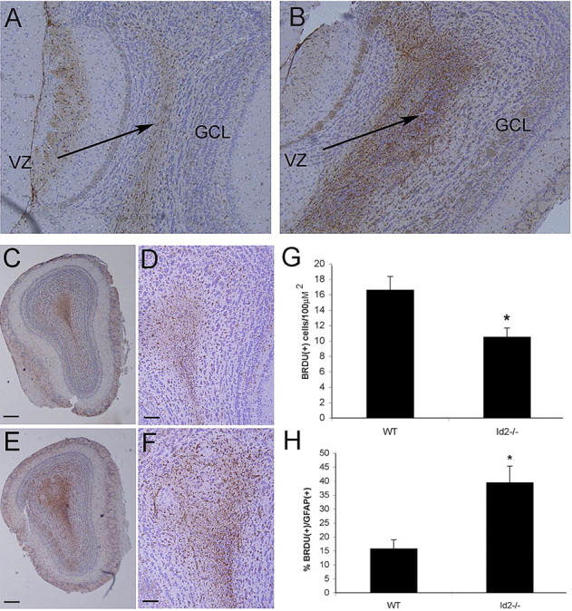
The Id2−/− olfactory bulb has increased numbers of newborn astrocytes. GFAP immunohistochemical analysis (brown staining) of sagittal (A, B) and coronal (C–F) histologic sections of WT (A, C, D) and Id2−/− (B, E, F) OB counterstained with hematoxylin. A, B Arrows denote VZ. Original magnification 5×. C, E, Original magnification 5×; scale bars, 100 μm. D, F, Original magnification 20×; scale bars, 50 μm. G, Using immunofluorescence on frozen sections from animals inoculated for 7 d with BrdU, total BrdU-positive cells were quantified in serial confocal micrographs taken from the OB of WT and Id2−/− mice. H, BrdU/GFAP double-labeled newborn astrocytes were quantified in multiple fields (n = 36 fields per genotype) from serial tissue sections and expressed as a percentage of total cells. Error bars represent SEM (E) p < 0.023. F, p < 0.005.
