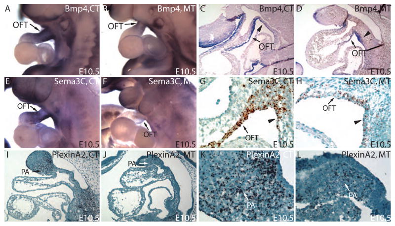
Figure 6. Defective OFT myocardial accruement of SHF mesodermal cells disrupts the normal distribution of signaling molecules essential for NCCs migration.
(A,B) Whole-mount RNA in situ hybridization revealed a reduction in Bmp4 expression in OFT. (C,D) Close examination of the subsequent sections further revealed that the reduction of Bmp4 expression is in OFT myocardium. (E,F) Whole-mount in situ hybridization reveals a marked reduction in Sema3C in OFT cuff in mutant compared to the control. (G,H) RNA In situ hybridization on paraffin section showed that the Sema3C was mainly expressed in outflow tract myocardium and was significantly down-regulated in mutants compared to the controls. (I,J) RNA In situ hybridization on paraffin section reveals a lower level of PlexinA2 transcripts in PA mesenchyme cells. K and L are higher magnification views of PA region in I and J. Triangle points to the splanchnic mesoderm within the SHF.

