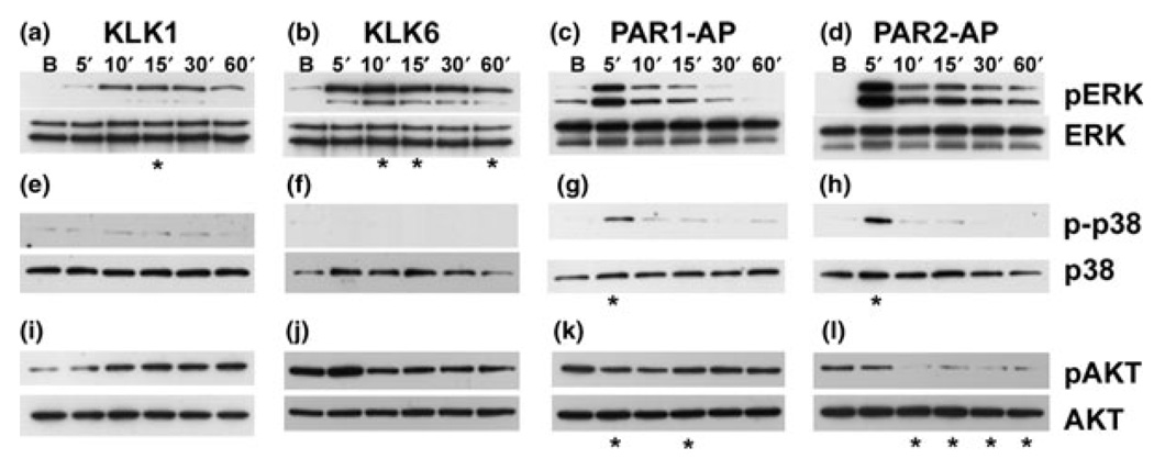
Fig. 8.
KLK1, KLK6, and PAR1- and PAR2-AP induced signaling in Neu7 astrocytes. Western blots for MAPK family members and AKT following exposure of Neu7 astrocytes to KLK1, KLK6, PAR1-, or PAR2-APs. ERK phosphorylation following exposure to (a) KLK1 (200 nM), (b) KLK6 (200 nM), (c) PAR1-AP (40 µM), or (d) PAR2-AP (200 µM). p38 phosphorylation following exposure to (e) KLK1 (200 nM), (f) KLK6 (200 nM), (g) PAR1-AP (40 µM), or (h) PAR2-AP (200 µM). AKT phosphorylation following exposure to (i) KLK1 (200 nM), (j) KLK6 (200 nM), (k) PAR1-AP (40 µM), or (l) PAR2-AP (200 µM); *p < 0.05.

