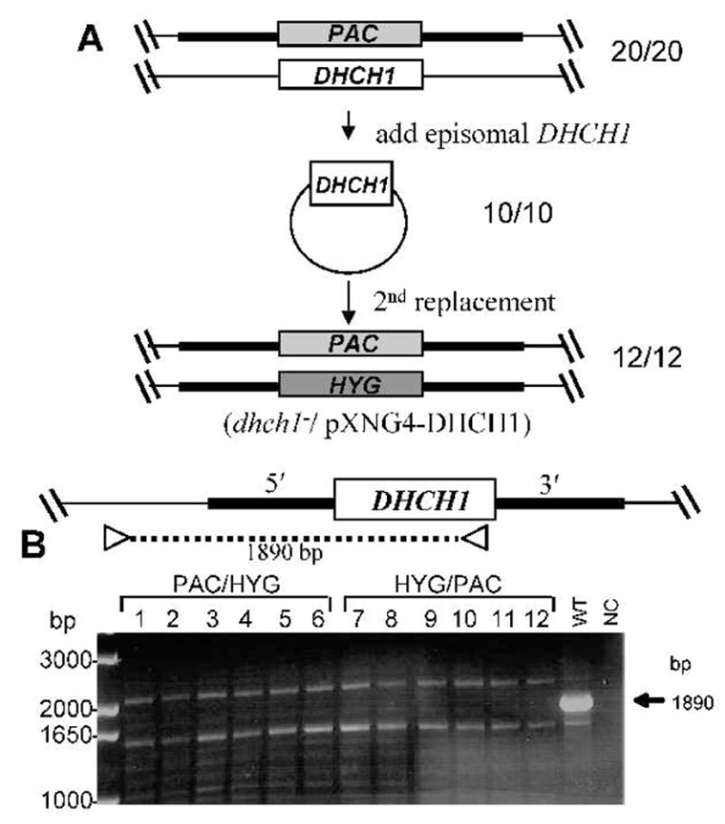
FIG. 3. Deletion of chromosomal DHCH1 in the presence of ectopic DHCH1.

A. Scheme for the deletion of chromosomal DHCH1 in the presence of ectopically expressed DHCH1. First, a pXNG4-DHCH1 episomal expression construct was transfected into a DHCH1/Δdhch1::PAC heterozygote described in Fig. 2A; 10/10 transfectants analyzed were confirmed to have the predicted genotype depicted. These DHCH1/Δdhch1::PAC [pXNG4-DHCH1] lines were then transfected with the HYG DHCH targeting fragment and selected for resistance to all three antibiotics; of the 12 clonal lines tested, all showed the planned replacement of the chromosomal DHCH1 locus (formally, Δdhch1::HYG DHCH1/Δdhch1::PAC [pXNG4-DHCH1]).
B – PCR tests showing loss of the chromosomal DHCH1. To distinguish the ectopic DHCH1 from the chromosomal locus, DNA from candidate lines was subjected to PCR tests using a using a primer directed against sequences located with the DHCH1 ORFt, and a primer located on the DHCH1 chromosome outside of the targeting fragment on the 5′ side (SMB2731/3126,, Table S1). WT, wild-type DNA control; NC, negative control lacking any DNA template. The arrow marks the size of the predicted fragment found in WT DNA, which is absent in the chromosomal dhch1− knockouts.
