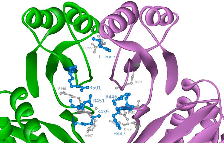
Figure 2. The Anion and Serine-Binding Sites of M. tb PGDH.
A close-up view of the anion and serine-binding sites depicted in Figure 1 is shown. At each interface between subunits A and B, there are two of each site related by approximate 180° symmetry. The view is down the axis connecting the two sites of the same type. One site is in front and is shown by blue ball and stick models. The second site is in back and is depicted by smaller gray ball and stick models. L-serine molecules are shown bound to the regulatory domain and the side chains of the cationic residues are shown in the anion-binding sites.

