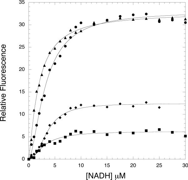
Figure 5. Fluorescence Resonance Energy Transfer in Native and Mutant M. tb PGDH.
Equimolar amounts of enzyme (2.5 μM subunit) were titrated with NADH in 200 mM potassium phosphate buffer, pH 7.5. The samples were excited at 295 nm and the fluorescence at 420 nm was monitored as a function of NADH concentration. Native, M. tb PGDH, • W29F, ▲; W327F, ■; and R451A-R501A-K439A,♦. The solid lines are fits to the Hill equation (Eq. 2).

