
FIGURE 1.
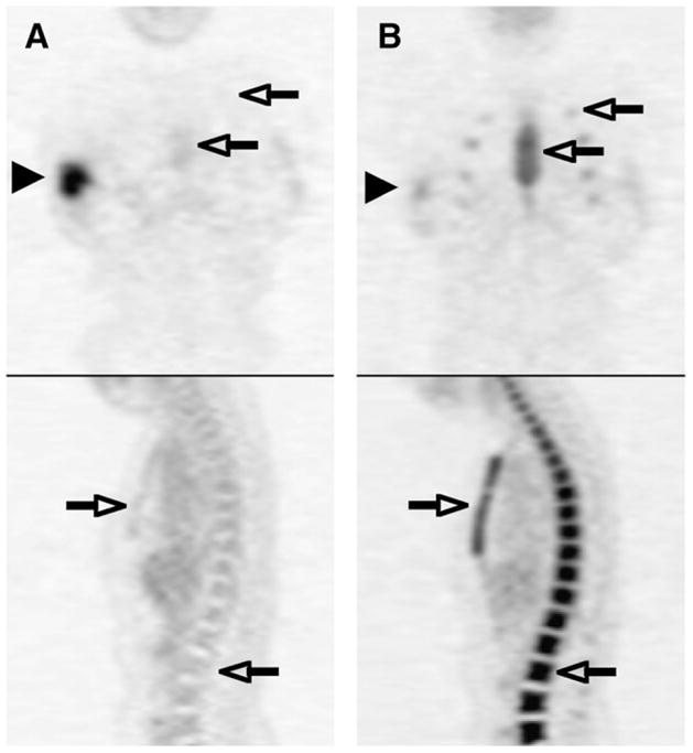
Coronal and sagittal 18F-FDG PET images of same patient before treatment (A) and after neoadjuvant chemotherapy including granulocyte CSF (B). Right breast cancer decreased in 18F-FDG activity after treatment (arrowheads), whereas 18F-FDG activity increased in marrow in spine, sternum, and ribs (open arrows).