Fig. 1.

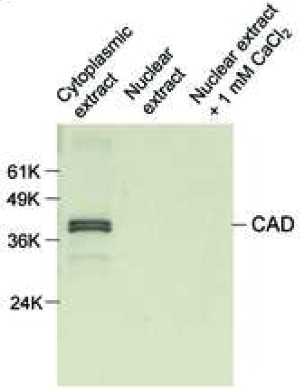
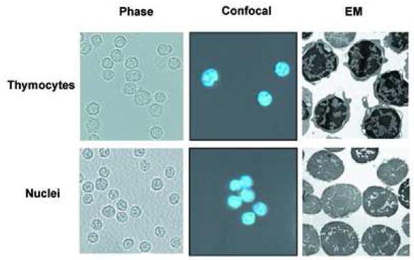
Morphological observation and the detection of CAD in nuclei isolated from viable rat thymocytes. (a) Thymocytes and isolated nuclei were obtained as described in Materials and Methods. They were observed by phase contrast microscopy (left panel; (Magnification, 200X), confocal microscopy (middle panel) or electron microscope (right panel, Magnification, 5,400X). The pictures of the nuclei indicate that cell membrane and cytoplasm are completely removed. Figures of phase contrast and electron microscopical analyses other than conforcal microscopy represent one of three independent experiments. (b) Isolated nuclei do not contain CAD. Fifty ug of cytosolic and nuclear proteins were analyzed by western blot with anti-CAD antibody. The data shown represents one of four independent experiments.

