Fig. 2.


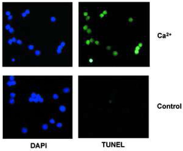
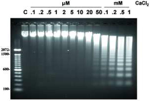
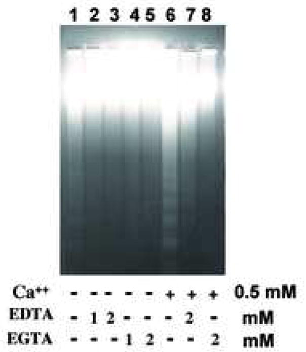
Ca2+ induces DNA cleavage and histone release in isolated nuclei from non-apoptotic thymocytes. (a) Induction of DNA cleavage in nuclei with 0.5 mM CaCl2 treatment. 1× 10⁴ nuclei were treated with a fluorometric TUNEL assay (right) and counter stained with DAPI (left). The data shown represents one of three independent experiments. (b) Induction of DNA degradation with various concentrations of CaCl2 ranging from 0.1 μM to 1 mM. C is a control. Nuclei (2 × 10⁶) were incubated in vitro at 37°C for 90 min with or without CaCl2 as described in Materials and Methods. Nuclei were harvested and treated with protease K and RNase. DNA (2 μg) from the nuclei was run on an agarose gel and stained with ethidium bromide. Typical internucleosomal DNA ladders were observed starting around 50 μM CaCl2. The data shown represents one of at lease three independent experiments. (c) Divalent cations and Ca2+ specific DNA degradation in isolated nuclei. EDTA and EGTA block Ca2+ dependent DNA degradation in nuclei obtained from thymocytes. The data shown represents one of two independent experiments.
