Fig. 3.


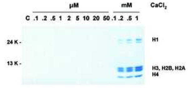
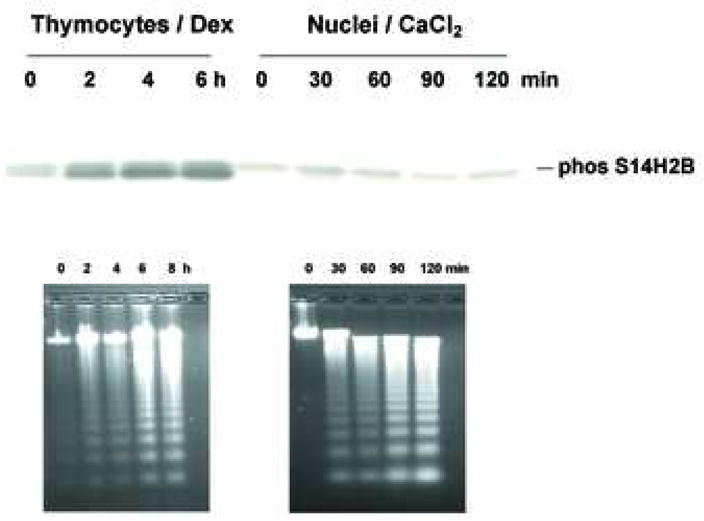
Histone release without H2B phosphorylation. (a) Histone release accompanying DNA fragmentation during incubation with CaCl2. After incubation of nuclei at each CaCl2 concentration as described for figure 2b, aliquots of lysis buffer soluble fractions were analyzed by SDS PAGE. Proteins were stained with Commasie blue. The data shown represents one of three independent experiments. (b) Ca2+ does not induce apoptosis-specific H2B phosphorylation in isolated nuclei. Western blot analysis with anti-phospho Ser14H2B for thymocytes cultured with dexamethasone or nuclei incubated with 1 mM of CaCl2 at 37°C for the indicated times. Histones were extracted were extracted from these samples and 10 μg were blotted against anti-phospho S14H2B. DNA was extracted and analyzed by agarose gel electrophoris. The data shown represents one of three independent experiments.
