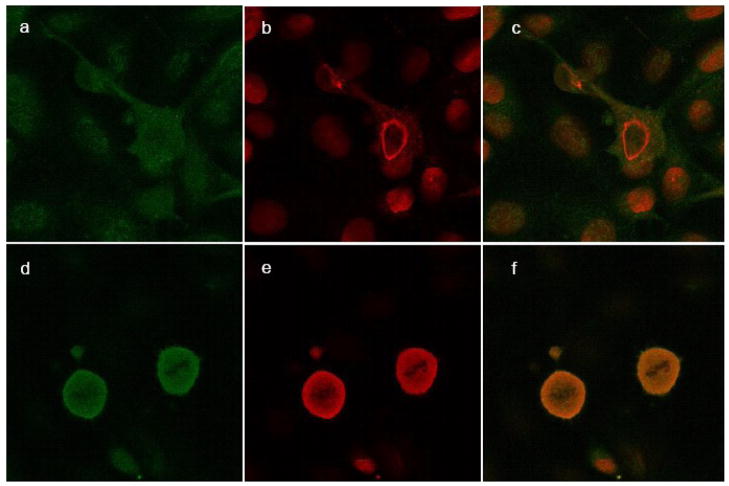
Figure 4.
Confocal microscopy of nuclear PLTP in SK-N-SH cells. SK-N-SH cells incubated without (a–c) and with LMB (d–f) for 24 hours were stained with monoclonal antibody against PLTP (green; a and d) and Lamin A, a nuclear marker (red; b and e); merged files (c and f). Secondary antibody controls were negative (not shown). The slides were observed under confocal Zeiss LSM 510 Meta microscope, using a water-immersion objective.

