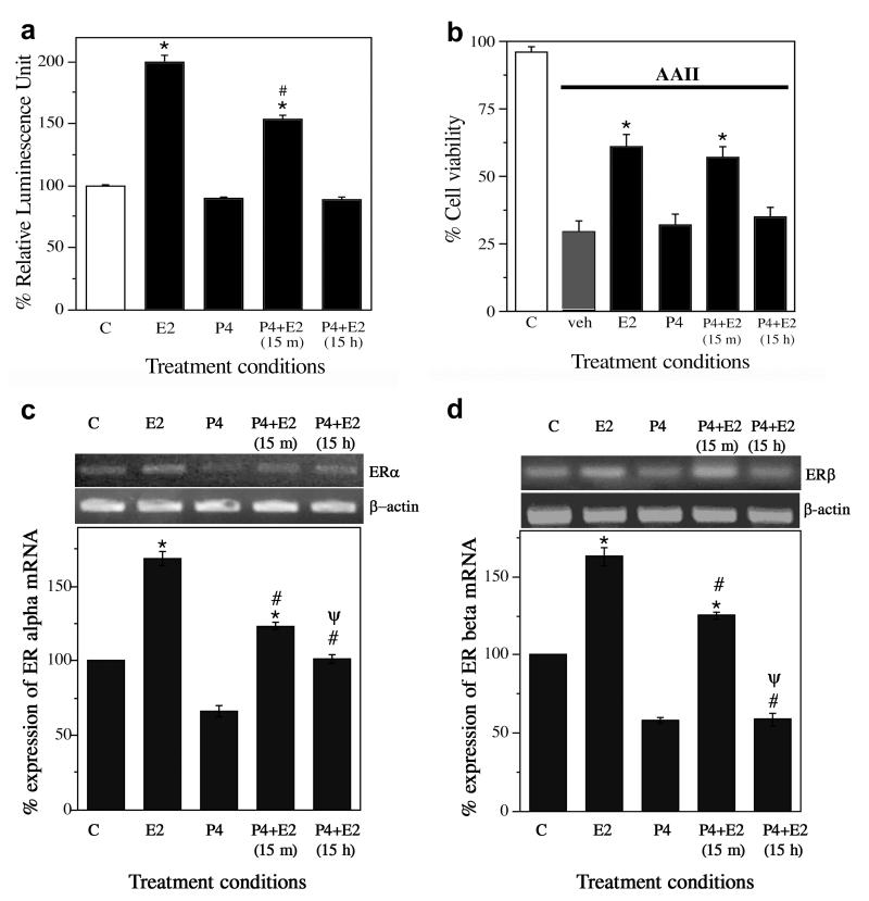
Figure 2.
Progesterone (P4) reduces oestrogen-induced increases in ER activity and neurone survival. (a) ER activity was determined by luciferase assay of neuronal cultures transfected with ERE-luc then treated with vehicle (C), 10 nM E2 (E2), 10 ng/mL (∼30 nM) (P4), or E2 with P4 pretreatment (P4+E2) for either 15 min or 15 h. Data show mean luciferase activity (±SEM) represented as relative luminescence unit and expressed as a percentage of vehicle-treated control condition. * Denotes p < 0.01 relative to vehicle-treated control (C, open bar) and # indicates p < 0.01 relative to E2 group. n=3. (b) Neurone survival was measured in cultures pretreated with 0 or 10 ng/mL P4 for 15 min or 15 h, followed by addition of 0 or 10 nM E2, and finally 24 h exposure to 3 μM apoptosis inducing peptide II (AAII). Cell viability data show average mean cell counts of viable cells (+SEM) expressed as percentage of vehicle-treated control group. * Denotes p < 0.01 relative to vehicle + AAII condition (veh, solid bar). n=5. (c, d) Levels of ERα and ERβ mRNA were determined under the same treatment conditions used in the luciferase assay. Data show representative agarose gels of RT-PCR products (upper panels) and mean (±SEM) mRNA levels determined by quantitative RT-PCR (lower panels). * Denotes increased expression (p < 0.0001) as compared to the vehicle control (C); # Denotes decreased expression (p < 0.0001) as compared to the E2 condition (E2); Ψ Denotes decreased expression (p < 0.001) as compared to the 15 m P4 pretreatment group [P4+E2(15 m)]. n=3.

