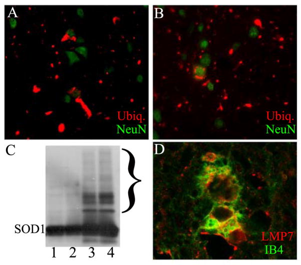
Fig 5.
Aggregate pathology in G93A/LMP2−/− mouse spinal cord. Immuno-staining of spinal cord from 7.5 month old G93A SOD (A) or G93A SOD1/LMP2−/− (B) mice for ubiquitin (red) or the neuronal marker, NeuN (green). (C) Western blot of spinal cord extract from 1 month G93A SOD1 (1), 1 month G93A SOD1/LMP2−/− (2), 7.5 month G93A SOD1 (3) or 7.5 month G93A SOD1/LMP2−/− mice probed with antibody to SOD1. Bracket highlights area of high molecular weight SOD1 positive complexes. 20 μg of protein loaded per lane. (D) Confocal microscopy showing immuno-staining for LMP7 (red) and IB4 lectin (green) within a microglial cell from the spinal cord ventral horn of 7.5 old G93A SOD1/LMP2−/− mouse. Magnification = 400× (A and B); 600× for D.

