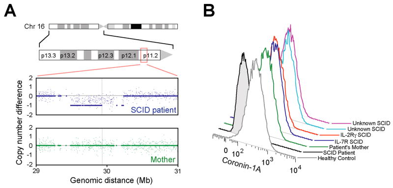
Figure 2.
A. Copy number analysis of patient and mother’s genomic DNA across a 2 Mb region of chromosome 16p11.2. Signal intensities from arrays hybridized to DNA from patient (blue) and mother (green) are normalized reference healthy control subjects.
B. Expression of Coronin-1A protein detected by fixation, permeabilizaton and intracellular staining of EBV transformed cells followed by flow cytometry.

