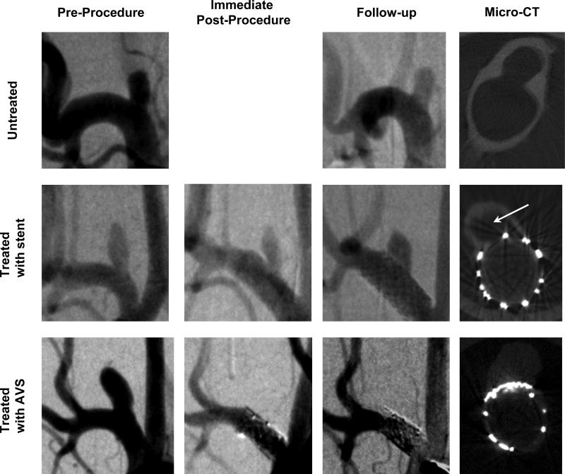
Figure 2.
Angiograms of aneurysms and Micro CT scans of the explanted aneurysms: untreated (subject #2 top row), treated with stent (subject #6 middle row) and treated with AVS (subject #11 bottom row). First three columns contain one angiographic instance: pre-procedure, immediate post-procedure and follow-up. Fourth column shows the central micro-CT slices taken perpendicular onto the aneurysm neck and parent vessel.

