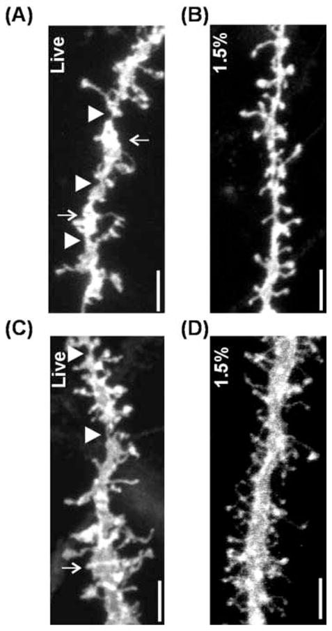
Figure 2.

Confocal microscopic images of dendritic segments. A,B, basal dendrites. C,D, apical dendrites. Images of dendritic segment from live slices (A,C) shows aberrant structures such as notching (arrow heads) and swelling (arrows). In contrast, dendritic processes from lightly fixed slices seems to be well preserved (B,D). Scale bar = 5 μm
