
Figure 4.
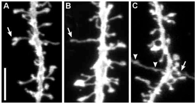
Special morphologies encountered in spine images from lightly fixed slices. A, branched dendritic spine. B, filopodial protrusion. C, A synaptic contact between an axon and a dendritic spine was accidentally imaged (arrow). Arrow heads represent boutons en passant of an accidentally stained axon. Scale bar = 5 μm