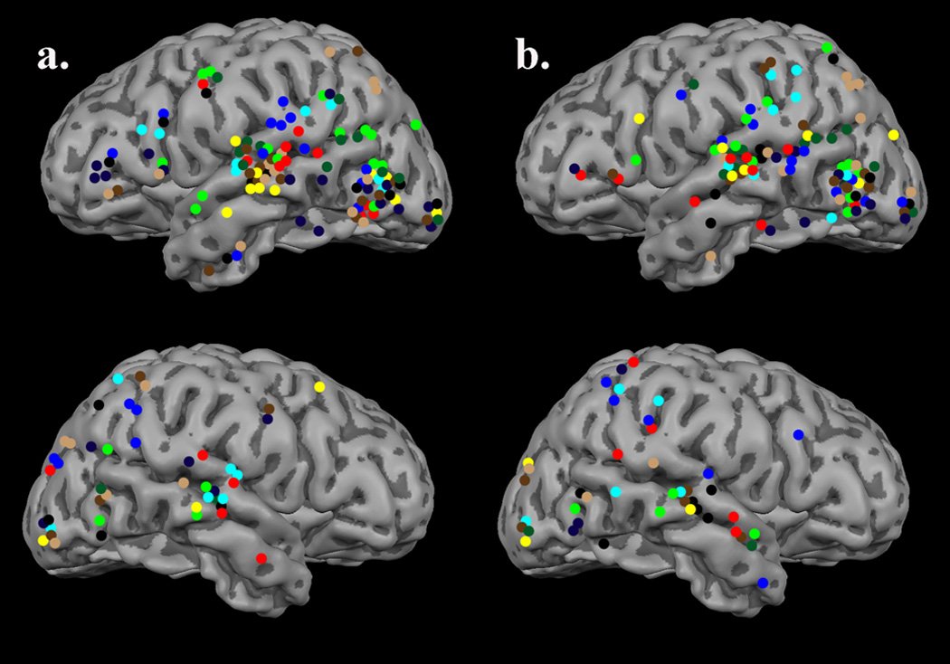
Figure 1.

Sources active 125–600 ms post-stimulus onset, in all subjects, have been projected to the surface of a standardized 3-D rendering of a participant’s MRI for easier visualization. The different colors represent different participants (i.e., all sources detected in the same participant are the same color). Sources in the (a) word condition and (b) pseudoword condition were strongly left-lateralized, and displayed remarkable spatial consistency within-subject. Each condition evoked substantial activation in the entire network of classically-recognized left hemisphere language processing areas. As shown, activation tended to cluster in posterior fusiform gyri, LOT/anterior fusiform region, left superior temporal areas, and to a lesser extent parietal regions. In some subjects, substantial activation was also present in the left inferior frontal gyrus.
