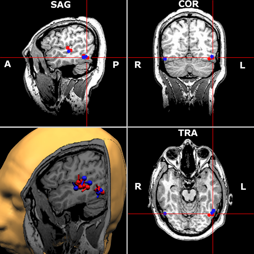
Figure 2.
Representative subject. Word elicited sources are depicted in red and pseudoword responses in blue. Both conditions evoked robust activity in a network of left hemisphere regions, including the superior temporal gyrus/sulcus and LOT cortices. Right occipitotemporal areas were also activated in this subject. As shown, source areas across the two conditions overlapped almost entirely. Right hemisphere sources were also detected (temporal lobe, not shown), but were far less numerous. The 2-D MRI plots are shown in radiological convention, and the cylindrical bar on each ECD (3-D rendition only) represents the orientation of the cortical current.

