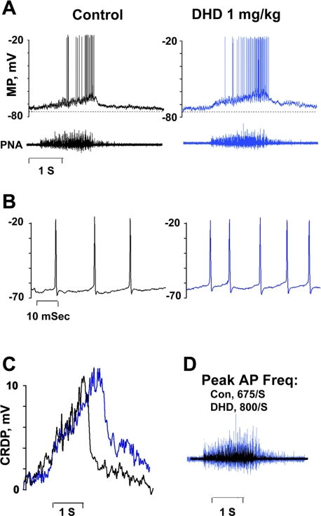
Fig. 2.
Effects of DHD given intravenously on a nonantidromically activated inspiratory neuron in the ventral medullary respiratory column (VRC). A: records of membrane potential (MP) and PNA electroneurograms before (left) and 2 min after injection of DHD (right). B: AP discharges presented on an expanded time scale, showing responses before (black, left) and after (blue, right) DHD. Discharges occurred at peak MP depolarization. C: central respiratory drive potentials (CRDP) recorded before (black) and after (blue) administration of DHD. CRDPs were extracted from the records in A by binomial smoothing: 30,000 iterations in each trace. D: superimposed PNA traces before (black) and after (blue) DHD. The blue trace is enlarged to reveal differences in temporal patterns of discharge. Con, control.

