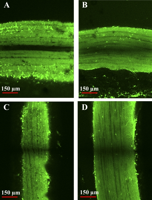
Fig. 10.
Immediate postbleach images of dark (A and C) and light (B and D) fibers during fluorescence recovery after photobleaching (FRAP). Fibers were incubated in the membrane permeable-dye calcein, which fluoresces green when hydrolyzed by intracellular esterases, and then subjected to a series of high-intensity bleach treatments. A and B: dark and light fiber postbleach images, respectively, from a FRAP experiment measuring radial diffusion coefficients. In dark fibers, calcein fluorophore (green) is thoroughly bleached within a single subdivision, and there is no radial diffusion into this bleached region, indicating cytoplasmic isolation. Light fibers exhibit some postbleach recovery via radial diffusion, indicative of cytoplasmic continuity throughout the fiber. C and D: images from measurements of axial diffusion coefficients. Pattern of recovery in the bleached region is similar between the dark (C) and light (D) fibers, indicating unhindered cytoplasmic exchange along the longitudinal axis in both fiber types. Subsequent images (not shown) demonstrate complete recovery of fluorescence in the bleached region.

