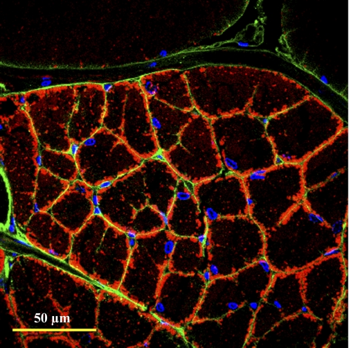
Fig. 8.
Aerobic dark fiber organelle distribution and perfusion. Transverse section of fibers from WGA-injected animals [to indicate perfusion pathways (green)] labeled for nuclei (blue) with 4′,6-diamidino-2-phenylindole and labeled for mitochondria (red) with MitoTracker Deep-Red 633. Nuclei are found exclusively at the subdivision edges and mitochondria at the edge and core of each subdivision. Intrafiber perfusion is indicated by complete WGA staining around each individual subdivision. Pattern is the same in small and large fibers.

