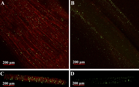
Fig. 9.
Pattern of hemolymph perfusion of dark (A and C) and light (B and D) levator fibers. Live animals were injected with a red-fluorescent Alexa 594-conjugated WGA and 0.2-μm yellow-green fluorescent microspheres. WGA binds to sarcolemmal (and vessel endothelial) glycoproteins, whereas microspheres become lodged in the smallest microvasculature of the muscle. A and B: 3-dimensional reconstructions (stacks) of whole fiber bundles. C and D: digitally reconstructed images of A and B, respectively, viewed in cross section. Fibers appear slightly flattened because of pressure from the coverslip. Intense WGA staining and substantial accumulation of microspheres inside the dark fibers indicate a high degree of intrafiber perfusion. Light fibers, however, exhibit very faint WGA staining and a lower abundance of microspheres (B and D), both of which occur primarily at the fiber edge. Beads that appear to be lodged inside the light fibers are likely located within fiber clefts.

