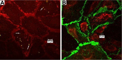
Fig. 1.
Invasion of Francisella tularensis live vaccine strain (Ft LVS) into an intact pulmonary microvascular endothelial cell (PMVEC) monolayer. Ft LVS were incubated at the abluminal surface of the PMVEC monolayer [5 or 500 multiplicity of infection (MOI)] for 6 h; then monolayers were collected and fixed for microscopy. A: at ∼5 MOI, staining with rabbit anti-Ft LVS antibody (arrows) and secondary FITC reveals individual Ft LVS and staining with anti-platelet endothelial cell adhesion molecule (PECAM) antibody and secondary Texas Red reveals intercellular junctions. B: at 500 MOI, staining with rabbit anti-Ft LVS antibody and secondary Texas Red and staining with anti-PECAM antibody and secondary FITC reveal numerous intracellular bacteria. Images represent sections from 4 experiments.

