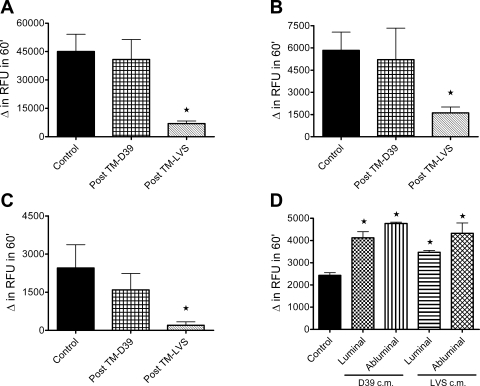
Fig. 6.
Elastase release from PMN after transendothelial migration (post TM). A–C: fluorometric analysis of elastase release from control PMN and PMN after transendothelial migration in response to S. pneumoniae strain D39 or Ft LVS. Elastase release after pretreatment with dihydrocytochalasin B followed by stimulation with bacterial chemoattractant formyl-Met-Leu-Phe (fMLF; A), stimulation with fMLF alone (B), or stimulation with PMA (C) was significantly inhibited in PMN that migrated across the endothelium in response to Ft LVS compared with control PMN or PMN after migration in response to D39. Values are means ± SE (n = 5). *P < 0.05 vs. control. D: elastase release in response to fMLF alone was enhanced in PMN incubated with bacteria-endothelial cell-conditioned medium (cm) for 3 h. This significant increase in elastase release occurred in response to D39- and Ft LVS-endothelial cell-conditioned medium from luminal and abluminal surfaces of the endothelial monolayer. Values are means ± SE (n = 4). *P < 0.05 vs. control.

