Fig. 3.
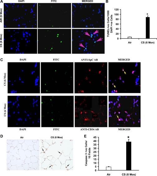
Chronic CS exposure causes alveolar cell apoptosis in A/J mice. A: lung sections (n = 5) from air- or CS-exposed (6-mo) mice were subjected to TUNEL (middle) and DAPI (left) staining. Merged images are shown at right. Overlapping DAPI in blue and FITC in green create a magenta, apoptotic-positive signal. B: CS-exposed mice (6-mo) showed abundant TUNEL-positive alveolar septal cells compared with air-exposed mice (n = 5 mice/group). Values are represented as means ± SE. P ≤ 0.05. C: identification of apoptotic type II epithelial cells (top) and endothelial cells (bottom) in the lungs of CS-exposed mice (6-mo). Type II epithelial cells and endothelial cells were detected using anti-SPC and anti-CD34 antibodies, respectively. Nuclei were detected with DAPI (blue). The merged images with colocalization of cell-specific markers (cytoplasmic red signal) with apoptosis (green FITC + blue DAPI) signal, resulting in lavender-like signal (yellow arrows), are shown. D: active caspase-3 expression in lung sections from chronic CS-exposed (6-mo) mice. CS-exposed A/J mice show increased numbers of caspase-3-positive cells (indicated by arrows) in the lungs (n = 5/group) (magnification, ×20). E: number of caspase-3-positive cells in the lungs of age-matched air- or CS-exposed mice. Caspase-3-positive cells were significantly (*P ≤ 0.05) higher in the lungs of 6-mo CS-exposed mice than the air-exposed mice. Values are represented as means ± SE.

