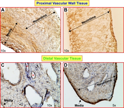
Fig. 3.
The structure and cellular composition of the occluded proximal pulmonary arteries and the vessels distal to the occlusion. Immunohistochemistry shows that a large amount of highly disorganized SM α-actin-positive (SM-αA+) cells (brown) in the proximal vascular wall (A and B). Tissue sections for A and B were prepared from 2 different patients. In the distal vascular tissue, the lumen was separated by the recanalizing fibrotic thrombus, and multiple channels (*) were observed in patients with CTEPH (C and D). Several medial layers of occluded vessels are shown in the proximal vascular tissue (A; labeled with “Media”) with strong SM-αA staining. The medial layer of the pulmonary vessel distal to the occlusion was clearly distinguished from neointima as a compact SM-αA+ cell layer (D). Medial and neointimal layers are indicated with arrows.

