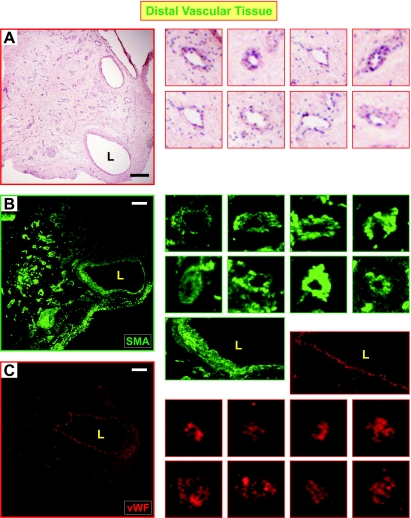
Fig. 4.
The recanalized regions in distal PTE tissues are surrounded by SM and endothelial cells. A: HE staining of a distal vascular tissue showed the lumen (L) and multiple-channel structure (as shown in images on the right), which was positive for SM-αA (B) and von Willebrand Factor (vWF; C) as shown by immunofluorescent staining. The images on the right are magnified channels from the panels on the left highlighting SM and endothelial cell staining in the distal pulmonary vessels. The 2 large images on the right in B and C are magnified areas around the lumen with SM-αA (green) and vWF (red) staining. The SM-αA and vWF panels are images from different sections obtained from the same region of the endarterectomized tissue; they are not overlaid images. Scale bar reflects 100 μm.

