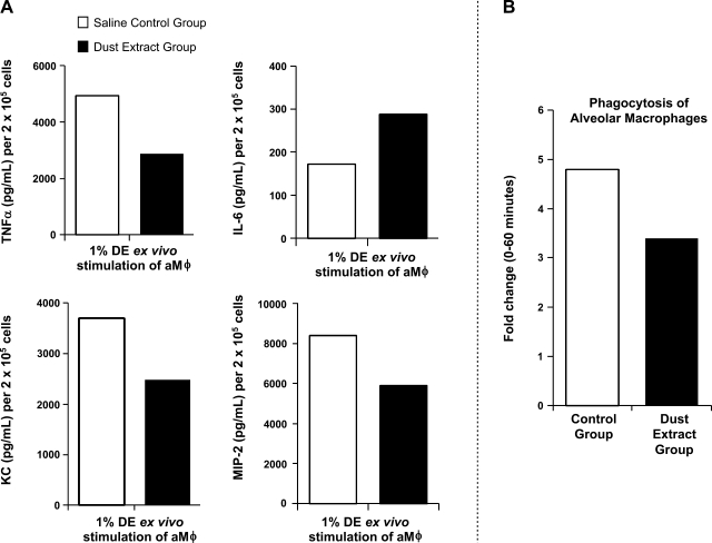
Fig. 4.
Ex vivo studies of alveolar macrophages (aMφ) from 2-wk saline control and DE (12.5%)-treated mouse groups (pooled aMφ; n = 8 mice/group). A: TNFα, IL-6, KC, and MIP-2 secretion in culture supernatant 5 h after stimulation with 1% DE. B: phagocytosis of IgG-opsonized Saccharomyces cerevisiae zymosan bioparticles with fold change in mean fluorescence intensity (proportion of cells in zymosan-exposed population at 60 min compared with cells exposed for 0 min).

