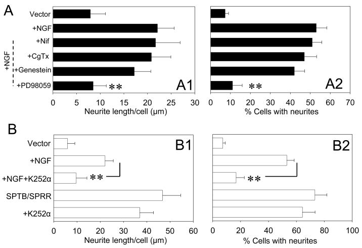
Figure 2.
NGF-induced neurite outgrowth is calcium independent. A. Quantification of neurite length (A1) and the percentage of cells with neuritis (A2) in cells expressing vector in the presence or absence of NGF with or without the indicated treatments after four day culture. NGF (50 ng/ml) and other drugs were added at the same time in the culture medium 3–4 hr after the cells were plated into 35 mm Petri dishes. Values are the mean ± SEM of the measurements made in four cultures in different days per treatment condition (100–150 cells were evaluated per treatment condition). *p<0.05, **p<0.01 compared to NGF- treated group, ANOVA followed by Holm-Sidak methods for multiple comparison. B. Quantification of neurite length (B1) and the percentage of cells with neuritis (B2) in cells expressing either vector or SPTB/SPRR Numb in the presence or absence of NGF in combination with or without K252α. Values are the mean ± SEM of the determinations made in four cultures in different days per treatment condition (100–150 cells were evaluated per treatment condition). *p<0.05, **p<0.01 compared to the NGF treated group, ANOVA followed by Holm-Sidak methods for multiple comparison.

