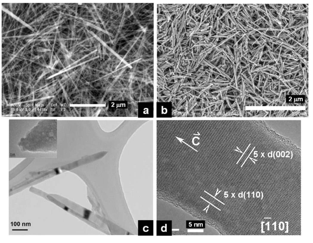
Figure 3.
(a) SEM image of long FA nanorods / nanowires prepared at pH 6.0 in the presence of EDTA and treated for 6 hours hydrothermally at a temperature of 121 °C and pressure of 2 atm; (b) SEM image of enamel crystals isolated from maturation stage of rat incisor enamel. The isolation methods see Chen et al., 2003 and 2005; (c) TEM image of the long needle-like synthetic FA nanorods / nanowires; (d) HRTEM image of the long synthetic FA nanorods / nanowires.

