Fig. 2.

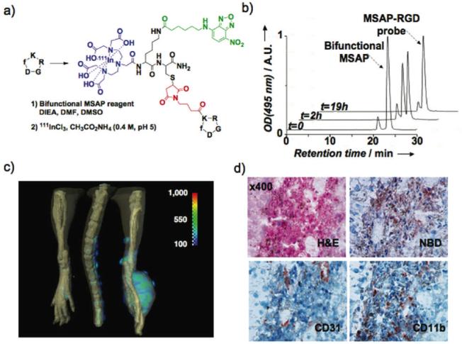
Molecular imaging with a bifunctional MSAP-RGD probe. a) The bifunctional MSAP (Scheme 1) was attached onto the lysine side chain of the cyclo[-RGDfK-] tumor-targeting peptide. b) The reaction between the bifunctional MSAP and the RGD substrate was monitored by RP-HPLC using NBD’s absorbance. c) After complexation with 111InCl3, the bifunctional MSAP-RGD probe was injected into a tumor-bearing mouse and monitored by SPECT-CT. d) Distribution of the probe within the tumor was visualized by immunohistochemistry with an antibody to the NBD hapten and compared to the distribution of CD31 (marker for endothelial cells) and CD11b (marker for monocytes/macrophages)
