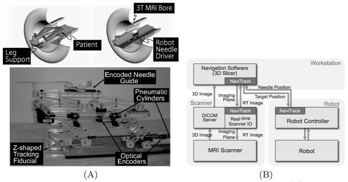
Fig. 1.
(A) A robot for transperineal prostate biopsy and treatment [9]. Pneumatic actuators and optical encoders allow operating the robot inside a closed-bore 3T MRI scanner. Z-shape fiducial frame was attached for a calibration. (B) The diagram shows communication data flow of the proposed software system.

