
Figure 2.
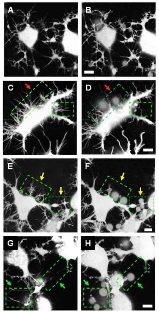
Morphological changes induced in neuronal cells by Aβ(1–42). Cells are loaded with calcein AM. Images A, C, E and G are taken as control before online addition of Aβ(1–42). Images B, D, F and H are taken 45 minutes after addition of Aβ(1–42). No morphological degeneration is observed for cells not treated with Aβ(1–42) (panels A and B). Significant degeneration is observed for cells treated with 50 nM Aβ(1–42) (panel D), indicated by arrows. Loss of cell-cell contact and neurite beading is observed after treatment with 500 nM Aβ(1–42) (panel F), and 5 μM Aβ(1–42) leads to loss of neuronal processes and fragmented neurites [11]. Scale bars 10 μm.