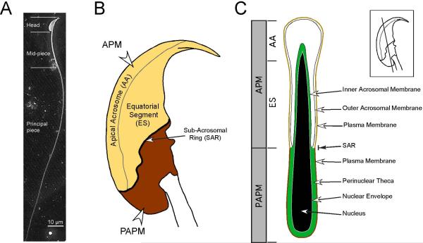
Fig. 1.
Organization of the membranes and structures of the murine sperm head. (A) Scanning electron micrograph showing the entire length of a murine sperm, including the falciform head and the flagellum. The flagellum is again divided into the midpiece, the principal piece, and a small terminal endpiece. (B) Schematic showing a lateral view of a murine sperm head. The plasma membrane of the sperm head is divided into two major regions, the APM and the PAPM, based on morphology and differences in membrane protein and lipid compositions. The APM and PAPM are delimited by a topographical feature known as the sub-acrosomal ring (SAR). On the basis of structure and function the APM is itself divided into the apical acrosome (AA), the region where membrane fusion will take place between the plasma membrane and the acrosomal vesicle, and the equatorial segment (ES). The hook-like structure is called the perforatorium. (C) Schematic showing a longitudinal section through the murine sperm head. Inset lateral view schematic of the sperm head shows the orientation of this longitudinal section. Areas corresponding to the APM (AA and ES) and PAPM are indicated. The nucleus (black) and nuclear membrane (grey) are surrounded by a cytoskeletal meshwork called the peri-nuclear theca (green). The acrosome (white) is seated over the apical region of the nucleus. The section of acrosomal membrane closest to the nucleus is the inner acrosomal membrane (IAM) and the acrosomal membrane apposing the plasma membrane is the outer acrosomal membrane (OAM). The plasma membrane is tightly wrapped around the structures of the sperm head and is closely associated with the OAM in the region forming the APM and the peri-nuclear theca in the PAPM.

