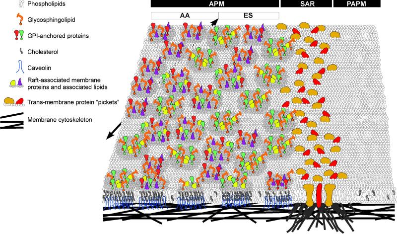
Fig. 11.
Schematic diagram showing a multi-level model for the mechanism of lipid segregation in the APM domain. (A) First level: there are free uni-molecular membrane lipids (e.g. most phospholipids) and proteins that follow the tenets of the Singer-Nicholson fluid-mosaic model (Singer and Nicolson, 1972). These can move across the SAR. (B) Second level: the existence of proteins and lipids in pre-assembled macromolecular composites. These could be heterogeneous or conform to specific compositional subtypes but are stable in not allowing sterol or GM1 exchange across the SAR. A less likely alternative at this level would be that the SAR could selectively prevent single molecules of specific lipid classes and a wide variety of proteins from diffusing across it, while allowing the movements described in the first level. (C) Third level: the existence of cytoskeletal-anchored transmembrane proteins as picket fences at the SAR restricts lateral diffusion of the macromolecular composites. This would require that all segregated molecules be components of such composites. Alternatively, cytoskeletal elements underlying the membrane might interact directly with membrane lipids and remove the necessity for transmembrane protein pickets. In this alternative, the pickets would be composed of the tethered, immobilized lipids. (D) Fourth level: spatial and temporal restrictions of specific sub-types of the macromolecular composites might lead to localized specializations (seen here distinguishing the regions of the AA and ES within the APM domain; a black arrow beneath the bilayer demarcates this region). Unidentified cytoskeletal proteins are shown interacting with the membrane at multiple locations, but in a more concentrated fashion at the SAR. Note: the depictions regarding the diversity of molecules are highly simplified in this schematic.

