
Fig.6. MHC II positive microglia from aged mice have a robust IL-1β induction following peripheral LPS injection.
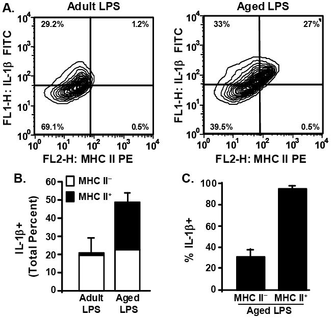
A subset of Percoll isolated microglia (Fig.5) were also stained with anti-MHC-II-PE. Cells were gated by CD11b+ staining. A) Representative bivariate contour plots of CD11b+ cells that were stained with anti-IL-1β-FITC and anti-MHC-II-PE. B) Average total percent of microglia that were IL-1β+ following peripheral LPS injection, differentiated based on whether they were MHC IIneg (white bars) or MHC II+ (black bars). Bars represent the mean ± SEM (n=3). C) Average percent of MHC IIneg and MHC II+ microglia that were also IL-1β+. Bars represent the mean ± SEM (n=3).