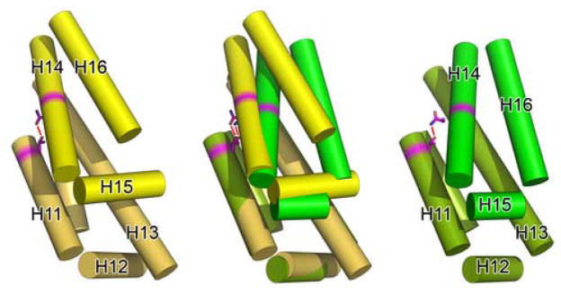
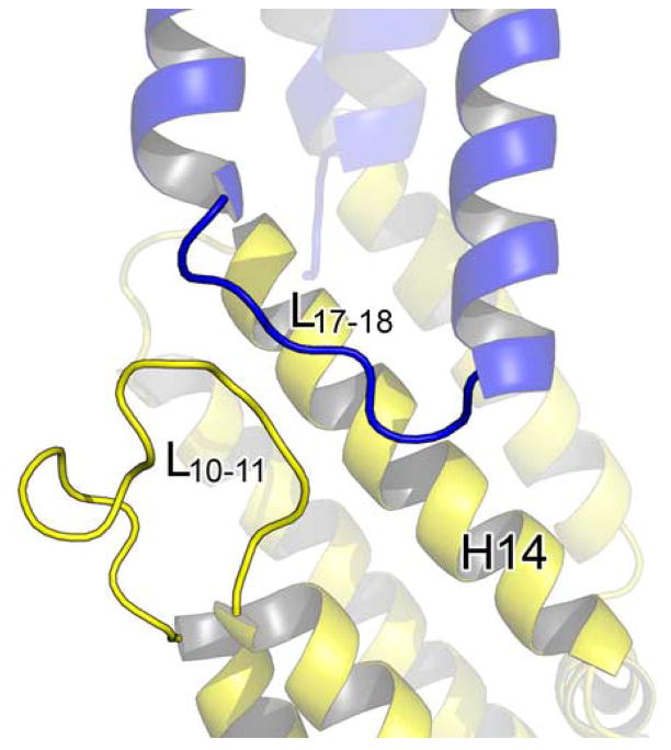
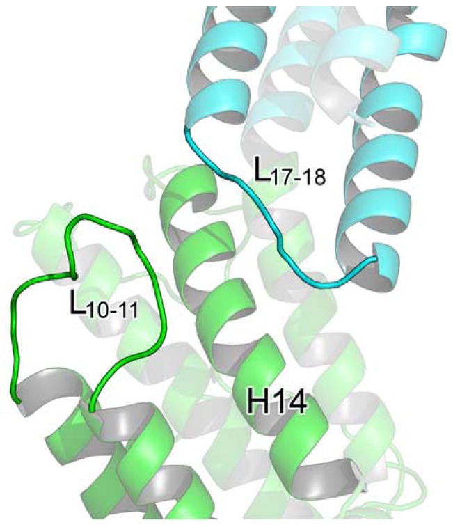
Figure 5. Differences in M domain packing affect M and C domain interactions.

(A) A cartoon diagram showing the alignment of MmExo70 with ScExo70 based on alignment of the N domain. Residues are colored by domain as in Figure 2. (B) Schematic diagrams of the M domains of Exo70 with helices shown as cylinders and all loops omitted. The left panel shows MmExo70 (yellow) and the right panel shows ScExo70 (green). H11-H16 are labeled. The middle panel shows the alignment of MmExo70 with ScExo70 based on H10-H13. The completely conserved hydrogen bond between MmExo70 Thr421/ScExo70 Thr372 and MmExo70 Asn498/ScExo70 Asn479 is shown as a red line. Note the differences in α-helical packing between the two molecules. (C) A cartoon diagram showing the positions of L10–11 and L17–18 in MmExo70. Relevant α-helices and loops are labeled. (D) A cartoon diagram showing the positions of L10–11 and L17–18 in ScExo70. Relevant α-helices and loops are labeled.


