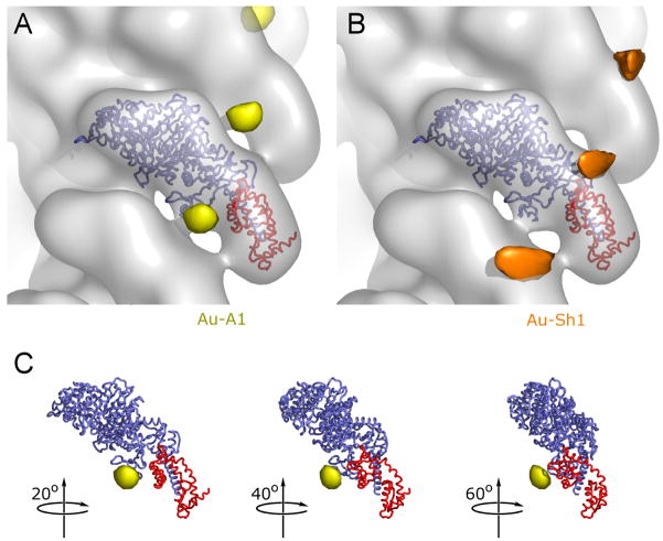
Figure 4.
Localization of the A1 extension by cryoEM. A reconstruction of actin decorated with S1 containing undecagold gold-labeled A1 (at –Cys3) is shown in transparent gray (pointed end up). The backbone of the docked crystal structure of chicken skeletal S1 is shown with the motor domain in blue and the ELC in red. The difference peak attributed to the A1 gold cluster label is shown in yellow (A and C); the difference peak attributed to the gold label on the reactive cysteine (SH1) of the heavy chain is shown in orange (B). This peak is located at the entrance of a narrow cleft between the converter/relay-loop and the rest of the myosin head, with the solvent exposed sulfhydryl group of SH1 as its bottom. This peak is approximately 10 times stronger than the peak in (A). Both contour levels were adjusted to optimize visibility.

