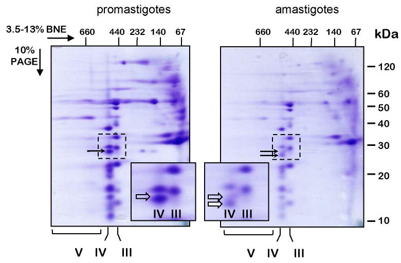
Fig. 3.

Separation of Leishmania donovani respiratory complexes in native gels. Mitochondria were isolated from promastigote and amastigote cells, lysed with 1% dodecyl maltoside and fractionated in 3.5-13% gradient Blue Native/10% Tris-tricine-SDS two-dimensional polyacrylamide gel. The gel was stained with Coomassie Brilliant Blue R250. Positions of the respiratory Complex V (ATP synthase), Complex IV (cytochrome c oxidase) and Complex III (cytochrome bc1) are shown below the gel. The inset panels represent the gel areas (bordered with a dotted line) containing an amastigote-specific band of Complex IV (filled arrow). The promastigote-specific subunit trCOVI, which is highly reduced in amastigotes, is shown with a transparent arrow. The native gel dimension was calibrated with an HMW Native Marker Kit (GE Healthcare) and the denaturing dimension with BenchMark™ Pre-stained Protein Ladder (Invitrogen). The size markers are shown above and to the right of the gel panels.
