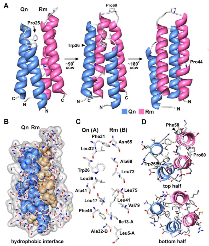
Figure 4.

The structure of an Rm-Qn four helix bundle.
A. A rotation series of the Rm-Qn 4-helix bundle is presented with the molecules displayed as ribbons. This structure is from the derivative crystals.
B. Non-polar side chains in the Rm-Qn interface are shown as solid spheres and are color coded for those from Qn (blue) and from Rm (tan).
C. Side chains in the Rm-Qn interface are shown as stick models with CPK colors and are labeled.
D. A top view is shown of the Rm-Qn 4-helix bundle with α-helices displayed as ribbons and side chains as CPK stick models. The figure was made with Chimera.
