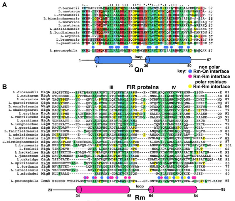
Figure 5.

Structure-based sequence alignments of the interacting regions of IcmQ and IcmR.
A. A sequence alignment is shown for Qn domains from 11 representative Legionella species and from C. burnetti. Hydrophobic residues are highlighted in green. The Qn α-helices (in blue) are aligned below the sequences and hydrophobic residues in the Rm-Qn interface are marked with blue dots.
B. A sequence alignment is shown for the middle regions of 24 FIR proteins. The Rm α-helices (red) are aligned with IcmR and FIR sequences. Residues in the Rm-Qn interface are marked with blue dots. Hydrophobic residues in the Rm-Rm interface are marked with red dots, polar residues are marked with yellow dots.
