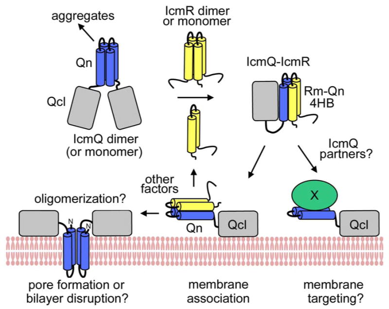
Figure 8.

A model for IcmR and IcmQ function.
(top left) IcmQ dimers are formed by pairwise interactions between the Qn domains and the resulting interface may be either parallel or anti-parallel (not shown). IcmQ also aggregates in the absence of IcmR which may be mediated by Qn. (top middle) IcmR may form a dimer using interactions between Rm regions in the absence of IcmQ. (top right) IcmR interacts with the Qn domain to form a 4-helix bundle which “disrupts” the IcmQ dimer and also dis-aggregates IcmQ. (bottom right) Possible IcmQ binding partners could displace IcmR and would potentially be targeted to the cell membrane by the Qcl region. (bottom middle) IcmR does not completely block membrane association of IcmQ which is mediated by binding of Qcl to the bilayer surface. (bottom left) IcmR must be displaced from IcmQ to allow Qn to interact with the membrane.
