
Figure 1.
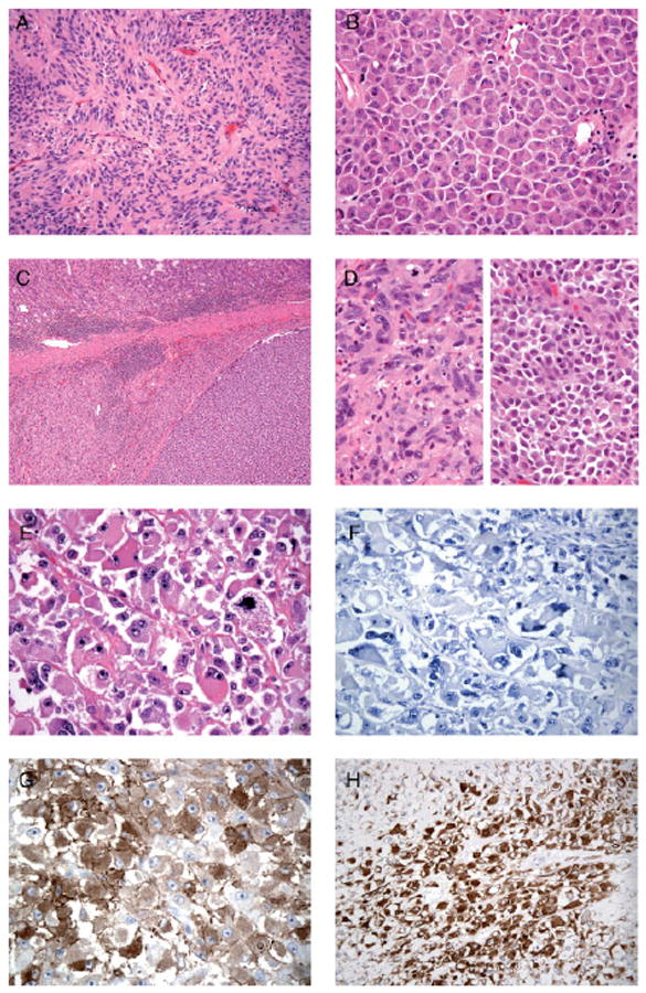
(A) GIST with spindle cell morphology composed of cells with a pale eosinophilic fibrillary cytoplasm, ovoid nuclei and ill-defined cell borders, with syncytial appearance and palisading (patient 3). (B) GIST with epithelioid cell morphology composed of round cells with eosinophilic cytoplasm arranged in sheets (patient 2). (C) Low-power view of a GIST in the stomach wall, with abrupt transition between two morphologically different tumour components (patient 14; KIT and PDGFR wild-type). Higher-power images of this tumour are demonstrated in (D), showing epithelioid morphology (right) and pleomorphic spindle cell morphology (left). (E) GIST showing unusual morphology, with huge epithelioid cells, vesicular nuclei and prominent nucleoli (patient 7; primary KIT exon 11 mutation). (F) KIT-negative GIST (patient 7). (G) KIT-negative GIST showing strong cytoplasmic staining for caldesmon (patient 7). (H) KIT-negative GIST showing strong cytoplasmic staining for desmin (patient 7)