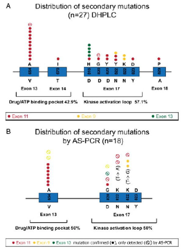
Figure 2.

(A) Summary of secondary KIT resistance mutations detected by D-HPLC in 27/57 tumour samples from 14 patients with progressing GISTs. Associated primary KIT mutations are indicated in red (exon 11 mutations), yellow (exon 9 mutations) and green (exon 13 mutations). (B) Summary of secondary KIT resistance mutations detected by AS–PCR only (
 ) or detected by both AS–PCR and D-HPLC (●) in 18 tumour samples. Associated primary KIT mutations are indicated in red (exon 11 mutations), yellow (exon 9 mutations) and green (exon 13 mutations)
